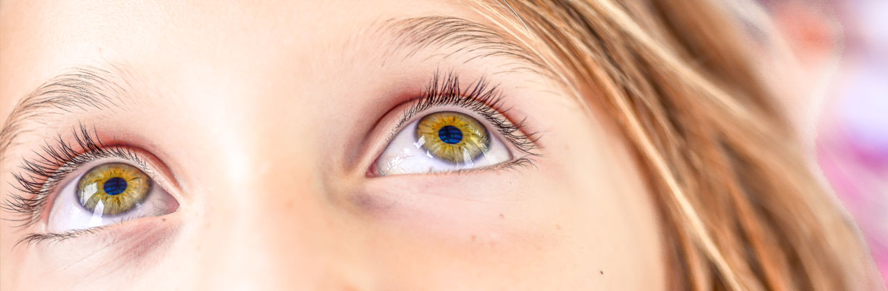
Iris Imaging: a closer look
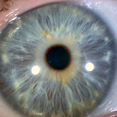
An iridology examination involves taking a very close look at the iris of each eye. A high resolution digital photo is taken of the iris, magnified and interpreted (this is called iris imaging). The image of the iris is projected onto a screen and the intricate details of the iris are made clearer and more visible. You can obtain copies of your iris imaging with an overview from your Sydney Iridologist. See: Iris Reports
Iris Imaging: Iridology Case Studies
How your iris appears is predominantly inherited from your ancestors. Even though the iris does take a long time to change, iris imaging gives you the opportunity to periodically monitor these changes.
Iris imaging is a close-up look at the iris signs. Careful and therapeutic interpretation of iris signs and their imaging can reveal health indications and genetic predispositions. See: Iris Signs
Iris imaging does not replace medical investigation and diagnosis. Iridology reveals what your health potential is and may guide you towards healthier lifestyle choices and therapeutic treatments. See: Naturopathy
If you have previously seen another iridologist and have earlier iris imaging, bring those photos with you when you come for your Sydney Iridology Consultation
Iris imaging iridology case studies are presented here:
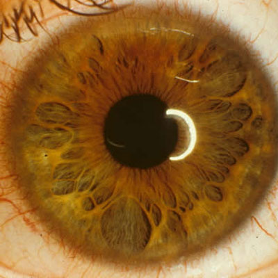
01: Digestive/Endocrine Systems
Iris imaging: Flower petal iris with hormonal indicators
A ‘flower petal’ iris may indicate a genetic predisposition to hormonal, endocrine and digestive weakness. The most common symptoms are irregular blood sugar levels, sudden mood swings, rapid exhaustion and an inconsistent digestive system function. Good digestive support, a diet full of natural nutrients and appropriate down time will be vital for this person’s general health and wellbeing.
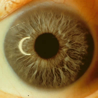
02: Inflammation
Iris imaging: Grey iris with rheumatic signs
A grey iris is relatively uncommon in this day and age and it tends to be observed in Northern European people. It can indicate a genetic predisposition to suffer from rheumatism, arthritis and skin conditions. This particular iris also has a white film super imposing the grey fibres that shows inflammation. Avoiding acidic-forming foods is very important at least by decreasing consumption of excessive coffee, alcohol, sweet products and grains. This person will benefit from an anti-inflammatory diet and lifestyle.
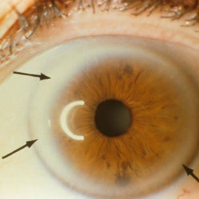
03: Cardiovascular/Digestive Systems
Iris imaging: Hardening of the arteries sign on brown iris
The thick white iris border as highlighted by the black arrows indicates a narrowing of the lumen of the arteries. This sign is commonly associated with high cholesterol and triglyceride levels in the blood and may also indicate a high LDL (the ‘bad cholesterol’) level and a low HDL (the ‘good cholesterol’) level. Psychologically, it can also indicate a strong determination in life when manifested in a positive form. When it’s manifested in its negative form, however, it can indicate a highly critical and demanding personality with a tendency toward self-righteousness. A diet rich in antioxidants will ensure that the blood purifies adequately and decreases the strong cardiovascular disease potential.
04: Digestive/Immune Systems
Iris imaging: Blue iris of a person later diagnosed with ulcers
The white bright halo centre is a sign of stomach inflammation. The dark orange and uneven pupil border indicates stomach malfunction. This person had many untreated stomach problems and was being treated symptomatically by the use of antacids such as Mylanta and Zantac. Despite this, vomiting, diarrhoea, severe stomach pain and reflux continued. The patient was later diagnosed with peptic ulcers and gastritis via endoscopy. A naturopathic treatment was prescribed with successful result.
The case study below demonstrates the importance of referring back to previous iris imaging.
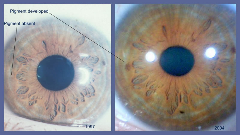
05: Fibroadenoma
Iris imaging: Right eye comparison showing brown spot pigment
The photo on the left was taken in 1997 and the photo on the right in 2004. The 2004 iris imaging shows a brown spot that was not visible in 1997. This spot is located in the right breast area. When this spot was observed in a 44 year-old female, Sydney Iridology referred her to a doctor for further examinations. She was subsequently diagnosed with a fibroadenoma (benign tumour) in her right breast and received appropriate treatment.
Not all brown spots mean tumours. Remember that it is important to observe the irises periodically and keep records of all images and changes. Changes in the iris can take years to develop and could indicate pathological conditions or tissue changes that will need to be medically investigated.
The iris imaging on this page is for case study purposes only.